What is the Normal Anatomy of the Foot and Ankle?
The foot and ankle form complex joints that are involved in movement and providing stability and balance to the body. The foot and ankle consist of 26 bones, 33 joints, and many muscles, tendons, and ligaments.
Bones of the Ankle
The ankle joint connects the leg with the foot and is composed of three bones: the tibia, fibula, and talus. The tibia or shinbone and fibula or calf bone are bones of the lower leg, which articulate with the talus or ankle bone, enabling up and down movement of the foot.
Three bony bumps present on the ends of the tibia and fibula form parts of the ankle joint:
- The medial malleolus, formed by the tibia, is found on the inside of the ankle.
- The posterior malleolus, also formed by the tibia, is found at the back of the ankle.
- The lateral malleolus, formed by the fibula, is found on the outer aspect of the ankle.
Bones of the Feet
The foot acts as a single functional unit, but can be divided into three parts: the hindfoot, midfoot and forefoot.
The hindfoot forms the ankle and heel, and is made up of the talus bone and calcaneus or heel bone. The heel bone is the largest bone in the foot.
The midfoot connects the hindfoot to the forefoot, and consists of one navicular bone, one cuboid bone, and three cuneiform bones. The navicular bone is found in front of the heel bone, and the cuneiform and cuboid bones are arranged in front of the navicular bone.
These bones are connected to five metatarsal bones of the forefoot that form the arch of the foot for shock absorption while walking or running. The forefoot is also made up of the toes or digits, formed by bones called phalanges - three in each toe, except the big toe, which has only two phalanges. The big toe has two additional tiny round sesamoid bones in the ball of the foot, which helps in upward and downward movements of the toe.
Ankle and Foot Joints
There are 33 joints in the ankle and foot. They include:
- Hinge joints in the ankle, which allow flexion (bending) and extension
- Gliding joints found in the hindfoot, which allow gliding movements
- Condyloid joints found in the forefoot and toes, which allow the flexion (bending) and extension, adduction, and abduction (sideward movement).
The joints of the foot and ankle provide stability and support the weight of your body, helping you to walk or run, and adapt to uneven grounds.
Soft Tissues of the Ankle and Foot
Our feet and ankle bones are held in place and supported by various soft tissues such as cartilage, ligaments, muscles, tendons, and bursae.
The joint surface of all the bones of the ankle and foot are lined by a thin, tough, flexible, and slippery surface called the articular cartilage, which acts as a shock absorber and cushion to reduce friction between the bones. The cartilage is lubricated by synovial fluid, which further enables smooth movement of the bones.
Ligaments are tough rope-like tissue that connect bones to other bones, and hold them in place, providing stability to the joints. The plantar fascia is the largest ligament in the foot, originating from the heel bone to the forefoot, it extends along the lower side of the foot and is involved in maintaining the arch of the foot. The plantar fascia ligament stretches and contracts to provide balance and strength to the foot. Lateral ligaments on the outside of the foot and medial ligaments on the inside of the foot provide stability and allow up and down movement of the foot.
The foot is made up of 20 muscles that help in movement. The main muscles include:
- Anterior tibial muscle, which allows up and down movement of the foot
- Posterior tibial muscle, which supports the arch
- Peroneal tibial muscle, which controls movement on the outside of the ankle
- Extensors, which enable the ankle to raise the toes just before stepping forward
- Flexors, which stabilize the toes against the floor
- Smaller muscles that help the toes to lift and curl
Tendons are soft tissues that connect muscles to bones. The largest and strongest tendon in the foot is the Achilles tendon, present at the back of the lower leg around the heel bone. Other tendons include peroneal and anterior and posterior tibialis.
Bursae are small fluid-filled sacs that decrease friction between tendons and bone or skin. They contain special cells called synovial cells that secrete a lubricating fluid.
-
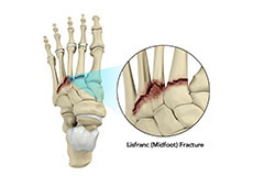
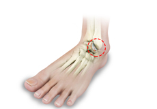
Lisfranc (Midfoot) Injury
The tarsometatarsal joint or Lisfranc joint is the region in the middle of the foot formed by the articulation of the tarsal bones (a cluster of seven bones) and metatarsal bones (a group of five long bones). This region supports the arch of the foot.
-

Ganglion and Soft Tissue Tumors
A ganglion is a round, sac-like swelling or a fluid-filled lump under the skin near your foot and ankle joint. It can become bigger or smaller over time and may be visible or not, especially if it is small.
-
Midfoot Arthritis
Midfoot arthritis is pain and inflammation of the midfoot. It occurs due to damage of cartilage or tissues around the joints. The damage may occur due to injury, aging or autoimmunity. The foot bones are the phalanges, the metaphalanges, and the tarsal bones. The midfoot consists of 5 bones called lesser tarsal bones. They are:
-
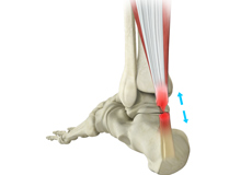
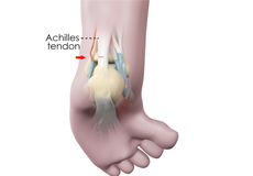
Achilles Tendon Rupture
The Achilles tendon is a strong fibrous cord present behind the ankle that connects the calf muscles to the heel bone. It is used when you walk, run and jump. The Achilles tendon ruptures most often in athletes participating in sports that involve running, pivoting and jumping. Recreational sports that may cause Achilles rupture include tennis, football, basketball, and gymnastics.
-
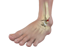
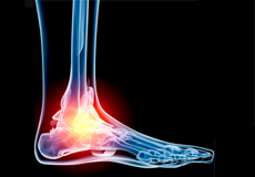
Ankle Fractures
The ankle joint is composed of three bones: the tibia, fibula, and talus, which articulate together. The ends of the fibula and tibia (lower leg bones) form the inner and outer malleolus, which are the bony protrusions of the ankle joint that you can feel and see on either side of the ankle.
-
Ankle Instability
The joints of the ankle are held in place and stabilized by strong bands of tissue called ligaments. Ankle instability is a chronic condition characterized by a recurrent slipping of the outer side of the ankle. It usually results from repeated ankle sprains, which are injuries to the ligaments. Ankle instability is generally noticed when you move your ankle joint but can also occur while standing.
-
Osteochondral Injuries of the Ankle
The ankle joint is formed by the articulation of the end of the tibia and fibula (shinbones) with the talus (heel bone). Osteochondral injuries, also called osteochondritis dissecans, are injuries to the talus bone. It is characterized by damage to the bone as well as the cartilage covering it. Sometimes, the lower end of the tibia or shinbone may also be affected.
-
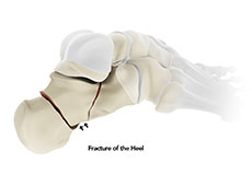
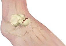
Heel Fractures
The calcaneus or heel bone is a large bone found at the rear of the foot. A heel fracture is a break in the heel bone due to trauma or various disease conditions.
-
Lisfranc (Midfoot) Fracture
The Lisfranc joint or tarsometatarsal joint refers to the region in the middle of the foot. It is a junction between the tarsal bones (bones in the foot arch) and metatarsal bones (five long bones in the foot). Lisfranc fractures can occur due to a fall from a height or a traumatic motor vehicle accident.
-
Talus Fractures
The talus is a small bone at the ankle joint that connects the heel bone and the shinbones, enabling up and down movement of the foot.
-
Foot and Ankle Trauma
The foot and ankle work together to provide balance, stability, movement, and propulsion to the human body. Its complex anatomy consists of 26 bones, 33 joints, muscles, tendons, ligaments, blood vessels, nerves, and soft tissues.
-
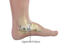
Ankle Ligament Injury
Ligaments are made up of elastic tissues that interconnect bones to one another. They bind the joint together, providing stability and support to the joint. The ligaments protect the ankle joint from abnormal rotation and stabilize the joint during movement.
-
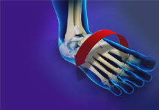
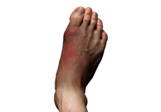
Foot and Ankle Arthritis
Arthritis is the inflammation of joints as a result of degeneration of the smooth cartilage that lines the ends of bones in a joint. This degeneration of the cartilages leads to painful rubbing of the bones, swelling, and stiffness in the joints, resulting in restricted movements.
-
Foot Infections
Foot infections may occur after trauma to the foot or loss of tissue because of contamination from foreign material and/or bacteria or fungus. Infections can occur in healthy individuals as well as in those whose health is compromised.
-
Congenital Limb Deformities
Limb deformities can be congenital (present at birth) or develop at a later stage because of a fracture, infection, arthritis or tumor.
-
Osteochondral Lesions of the Ankle
The tibia and the fibula bones of the lower leg join with the talus bone to form the ankle joint. The talus bone is an important bone located between the tibia and fibula and the heel bone (calcaneus).
-
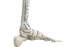
Internal and External Fixation of Foot and Ankle Fractures
Foot and ankle fractures are breaks or cracks in any bone of your foot and ankle joint. Fixation of fractures is a surgical method of reconnecting the broken or cracked bones and fixing them in the correct place using orthopedic hardware.
-
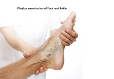
Foot and Ankle Examination
Foot and Ankle conditions typically occur due to injury of the muscles, ligaments or bones, due to aging, or certain disorders including gout, bunion, arthritis, claw toes, bursitis, hammertoes, stress fracture, etc.
-
Foot Rehabilitation Following Surgery
A foot injury or foot surgery may leave you immobile for a period of time. To return to your regular activities and more strenuous recreational activities, it is necessary for you to follow a well-planned activity and exercise program.
-
Foot Reconstruction
The foot is formed by several bones, ligaments, joints, and muscles that function collectively to control the various movements like walking and running. This complicated structure of the foot permits it to resist heavy forces every day.
-
Subtalar Arthrodesis
The subtalar joint is a complex joint located below the ankle joint and is formed by the union of the heel (calcaneus) and the talus (ankle) bone. The subtalar joint allows side-to-side movement of the foot.
-
Achilles Tendon Repair
Tendons are the soft tissues connecting muscle to bone. The Achilles tendon is the longest tendon in the body and is present behind the ankle, joining the calf muscles with the heel bone. Contraction of the calf muscles tightens the Achilles tendon and pulls the heel, enabling the foot and toe movements necessary for walking, running and jumping.
-
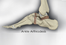
Ankle Arthrodesis
Ankle arthrodesis is recommended for the treatment of severe end-stage arthritis that has not responded to conservative treatment measures such as medications or injections. The other indications include ankle infections, neurological ankle instability, and tumors.
-
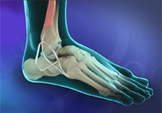
Ankle Ligament Reconstruction
A sprain is the stretching or tearing of a ligament. Ligaments connect adjacent bones in a joint and provide stability to the joint.
-
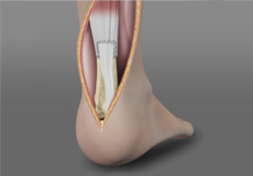
Ankle Tenotomy
Ankle tenotomy is a surgical procedure to lengthen the Achilles tendon, enabling the ankle to flex upward and allowing the heel to be placed flat on the floor. It is indicated in if you have an abnormally developed Achilles tendon or one that has become shortened and is difficult to stretch. The surgery is performed to restore the normal range of motion of the ankle.
-
Ankle Instability Surgery
Ankle instability is a chronic condition characterized by the recurrent slipping of the outer side of the ankle. Instability is generally noticed during movement of the ankle joint, but can also occur while standing.
-
Tendon Transfer
In a tendon transfer procedure, a healthy tendon is transferred to replace the damaged tendon and restore the normal movement of the foot.
-
Charcot Reconstruction
Charcot foot and ankle is a condition characterized by gradual weakening of the bones, joints and soft tissues, and loss of sensation in the foot and ankle. It is caused by nerve damage (neuropathy) in the foot and ankle or due to diabetes.
-
Foot & Ankle Deformity Correction
Foot and ankle deformity is the structural abnormality caused by misalignment of the bones of the foot and ankle.